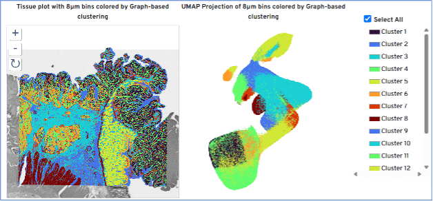
Molecular pathology is undergoing a revolution driven by spatial transcriptomics. By mapping whole transcriptome gene expression data onto intact tissue sections, it allows researchers to view cellular transcription in its true native microenvironment. This gene expression data can be overlayed with H&E or Immunofluorescence staining to visualise tissue morphology and protein expression from the same tissue section.
Visium technology:
Visium Spatial Gene Expression technology is engineered to map a tissue’s transcriptome directly onto a standard histological tissue section. The foundation relies on specialized glass slides covered with millions of arrayed oligo capture probes.
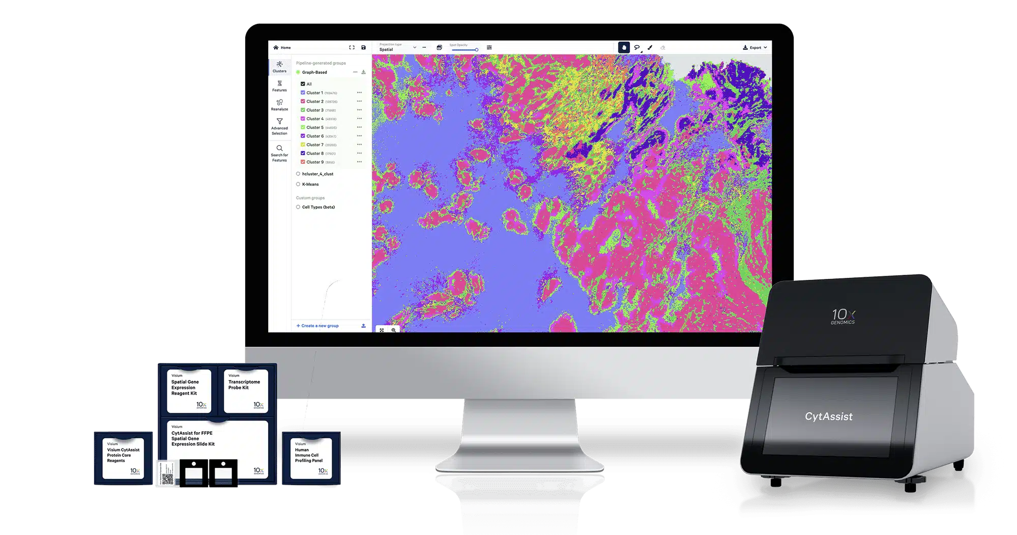
Historically, sectioning tissue directly onto specialised capture arrays has presented a processing bottleneck. The 10x Genomics Visium CytAssist platform bridges this gap, introducing a streamlined workflow that seamlessly couples standard clinical histology with high-definition spatial discovery.
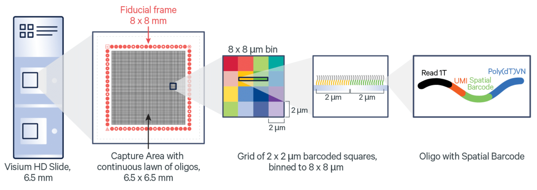
Each probe is built with unique architecture:
A Spatial Barcode that anchors every transcript to an exact X,Y coordinate on the glass surface.
A Unique Molecular Identifier (UMI) to allow for precise, de-duplicated transcript counting.
An Oligonucleotide Capture Sequence designed to capture specific transcripts or complementary cDNA.
When tissue is fixed, stained, and permeabilised on these slides, mRNA molecules (or hybridized probe pairs) release locally and bind to adjacent capture probes. The resulting libraries are sequenced via next-generation sequencing (NGS), and the barcodes map each gene expression profile back to its original location. This gives quantitative molecular resolution to morphology, identifying exactly which genes are active in specific tissue regions, immune niches, or structural boundaries.
The CytAssist Platform
The Visium CytAssist is a benchtop instrument which addresses the logistical constraints of direct-on-slide tissue sectioning. Traditionally, tissue sections had to be cut directly onto specialised, high-cost capture slides, which is a process with a high margin of operator error, section loss, or poor tissue placement.
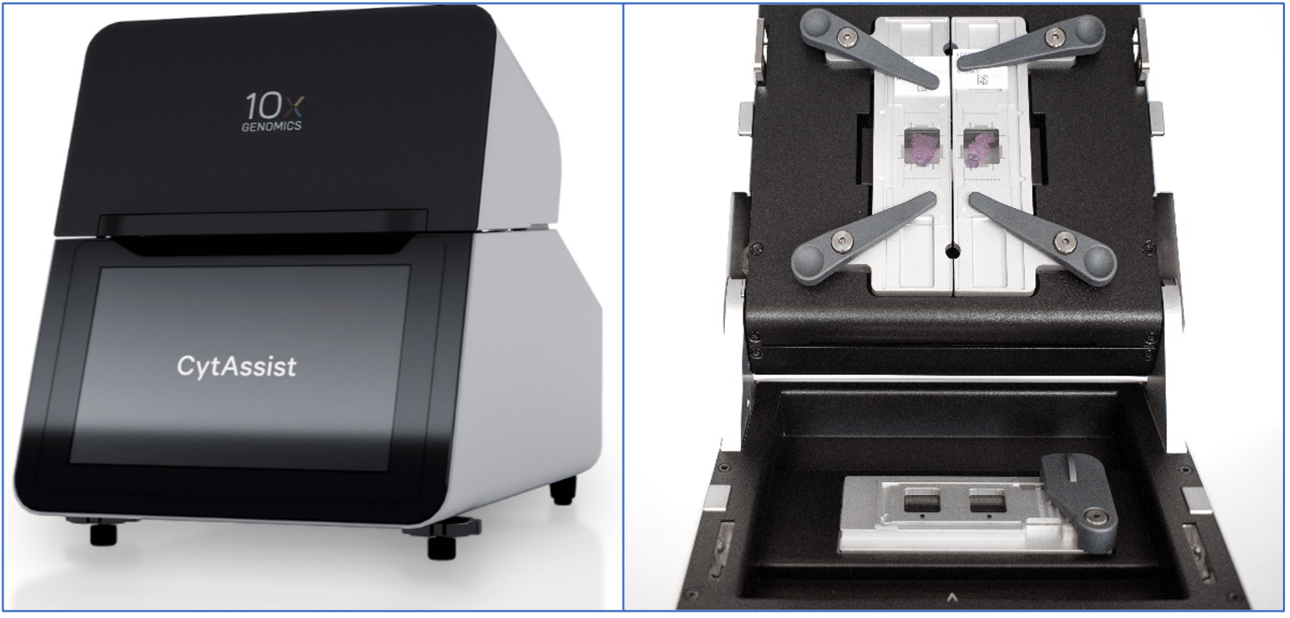
Rather than cutting tissue directly onto functional capture slides, tissues are sectioned, prepared, stained, and imaged on standard glass microscope slides first. The CytAssist instrument uses a microfluidic gasket assembly to align the standard tissue slide directly over a Visium capture slide.
By applying automated temperature control and precise reagent routing, it facilitates the migration of hybridized analyte molecules from the standard slide to the underlying Visium capture grid in under an hour.
This offers several advantages:
Archived samples: You can section tissue weeks in advance, pre-screen sections via H&E or immunofluorescence, and select only optimal, high-quality cuts for spatial running.
Precise Placement: You can preview exactly how the tissue fits over the array boundaries, completely eliminating off-target placement or tissue overlapping.
Clinical Sample Access: It opens up spatial transcriptomics to historical biobanks and clinical FFPE blocks that are difficult to cut directly onto specialised arrays.
Visium Slide Formats
To accommodate diverse tissue sizes and varying clinical needs, the Visium assay features two primary slide dimensions, both slide formats contain two capture areas per slide:
6.5 mm x 6.5 mm: Ideal for targeted biopsies, focused regions of interest (such as a core biopsy or a specific mouse brain hemisphere), and high-resolution exploratory assays where conserving sequencing depth per slide is vital.
11 mm x 11 mm: This layout expands the capture zone to provide an impressive 2.8x larger capture area. It is specifically designed for broader tissue samples, such as whole human organ resections, complex tumor-immune margins, or multi-tissue arrays.
The Visium/CytAssist Workflow
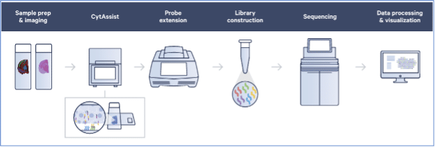
- Histology & Primary Imaging: Tissue blocks (FFPE or Frozen) are sectioned onto regular glass microscope slides. The sections undergo standard baking, deparaffinisation (if FFPE), staining (H&E or Immunofluorescence antibodies), and are imaged using a traditional high-resolution brightfield or fluorescence slide scanner.
- Probe Hybridization: For the probe-based assay (Visium HD WT Panel), a specialised probe pool targeting the entire transcriptome is added to the slide. These left and right probe pairs hybridize contiguously to their target mRNA sequences within the intact tissue and are ligated together to form a stable template.
- CytAssist Transfer: The standard glass slide is loaded into the CytAssist along with a functional Visium capture slide. The instrument takes an on-board image to catalog positioning, seals a precise microfluidic interface over the tissue, and uses a permeabilisation buffer to release the ligated probes, or native mRNA downward, capturing them onto the Visium slide’s spatial barcodes.
- Library Generation & NGS: The captured analytes are extended on the Visium slide surface to incorporate the coordinate barcodes and UMI sequences. The material is then collected, amplified via PCR, and prepared into an NGS library for sequencing.
Visium HD WT Panel vs Visium HD 3′ Assay
When designing your spatial experiments, it is vital to note that the Visium HD Whole Transcriptome (WT) Panel assay is carefully curated and exclusively validated for human, mouse, and rat species. If your research involves non-rodent model organisms, non-human primates, or other diverse species, you should instead utilise the Visium HD 3′ assay, which leverages an unbiased poly-A capture strategy to ensure robust transcript discovery across a significantly broader range of species. See table below for a comparison of the two Visium HD assays.
| Feature | Visium HD WT Panel | Visium HD 3' |
|---|---|---|
| Capture Strategy | Probe-Based Hybridization Employs highly curated, ligated probe pairs that target exact, predictable exonic sequences across the human, mouse or rat genome. | Poly-A RNA Capture Employs an unbiased oligo-dT surface capture mechanism that binds directly to the polyadenylated tails of active mRNA transcripts. |
| Tissue & Sample Compatibility | Highly Optimized for FFPE Superior performance on FFPE samples, archival tissues, or degraded clinical blocks where RNA may be fragmented. | Optimized for Fresh Frozen Designed for high-integrity, Fresh Frozen samples where intact, full-length transcripts are readily accessible. |
| Sensitivity & Data Specificity | High Specificity Because it relies on precise dual-probe ligation, it effectively filters out background artifact noise, off-target genomic DNA, and degraded fragments. | Broad Discovery Captures any polyadenylated molecule. This makes it highly advantageous for discovering novel splice variants, tracking somatic mutations, or mapping non-canonical transcripts. |
Visium Data Analysis
The transition from raw sequencing data to biological insights relies on robust, production-grade bioinformatics pipelines developed by 10x Genomics:
- Space Ranger Processing: The raw sequencing files (FASTQs) and the high-resolution tissue images are fed directly into the Space Ranger pipeline. The software processes the data by detecting the spatial barcodes, running strict quality filters, performing transcript alignment, and overlaying the matrix data cleanly onto the original histology image.
- Loupe Browser Visualisation: For an interactive data review, the output files load seamlessly into Loupe Browser. This intuitive desktop interface lets researchers easily toggle between the visual H&E morphology and gene expression heatmaps, manually select cellular regions of interest, and instantly isolate localised cluster markers.
- Open-Source Tools: For advanced bioinformatics and downstream modeling, Space Ranger matrices output standard h5 and matrix formats. These load directly into industry-standard open-source programming frameworks such as Seurat or Giotto in R, and Squidpy or Scanpy in Python, enabling spatial clustering, automated cell-type deconvolution, ligand-receptor interaction tracking, and spatial trajectory analysis.